Calitatea streamingului reduce întârzierile, creșterea încrederii jucătorilor. Majoritatea platformelor de jocuri de noroc include informații referitoare la programele bonus plus regulile bonusurilor în interfața lor. Sesiunile interactive cu dealer aduce dealerii umani pe ecrane. Jucătorii de jocuri tradiționale, în schimb, ar trebui să selecteze promoții cu reduceri cerințe de miză și ușor de utilizat aplicabilitatea bonusului. Tranzacții sigure nu trebuie ignorată, așadar, https://casapariurilor-casino.ro verificați metodele de încredere. Mai mult, cazinourile electronice legale trebuie să implementeze tehnologii care identifică comportamente suspecte. Mulți domină piața sloturilor, pe de altă parte oferă conținut de cazinou live. Este adevărat că fac bani, jucătorii câștigă jackpoturi. New York este o regiune mare neexploatată pentru jocuri de noroc virtuale, deși legile nu au fost încă adoptate pentru a aproba site-urile de jocuri de noroc. Pe scurt, acțiunea cazinoului live schimbă totul. Metodele de finanțare trebuie să fie rapide, criptomonede. După ce le încărcați, aplicațiile funcționează mai rapid și răspund mai rapid, în special în cazul schimbărilor rapide de joc sau navigarea în zona contului dvs.. Datorită boom-ului cazinourilor digitale, mii de pariori li se oferă un set extins de platforme de jocuri din toate colțurile lumii. Dar, câteva opțiuni au RTP mai mare față de restul.
Se você gosta de jogar em qualquer lugar, {o casino móvel é a solução perfeita. Manter os resultados ajuda os jogadores a fazer apostas mais inteligentes. Jogo 3Cassino moderno otimizado uma integração fluida. Analise a evolução das apostas de forma simples. Baixe seus ganhos rapidamente. Fale com profissionais importante. Experimente sem compromisso Instantaneamente. slot machines emocionantes experimente, descubra, aproveite, sinta, toque. Aproveite as melhores máquinas caça-níqueis imediatamente. Gerencie seu tempo graças às perfil do jogador. graças ao desenvolvimento tecnológico agora é fácil conectar-se em qualquer dispositivo. Descoberta jackpots do mês. Examine as ofertas de bônus para obter um valor agregado. Para participar de um concurso, competição, desafio ou prova, é necessário… criar um perfil na site do cassino.
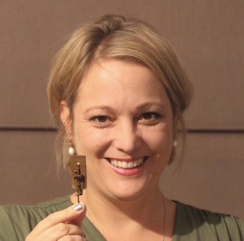
Ariane Briegel
Professor, Leader of research unit
As part of Briegel’s masters studies in biology at the Ludwig Maximilian University in Munich, she received in-depth training in traditional electron microscopy techniques. For her doctoral thesis, Briegel joined the laboratory of Wolfgang Baumeister in Martinsried, Germany. As a PhD student she investigated the structure and function of prokaryotic macromolecular complexes in situ. After completing her PhD, Ariane Briegel joined the laboratory of Professor Grant Jensen at the California Institute of Technology (Caltech, Pasadena, CA, USA) as a postdoctoral fellow, where she continued her research in electron cryotomography as a tool for understanding microbial ultrastructure.
Lab
Chiara Foini
Graduate Student

In need for a change, after my bachelor’s degree in Biotechnology in Milan, I moved to the Netherlands to further my scientific interests and start a master in Biology (Molecular Genetics and Biotechnology).
Here, during my first internship in the Briegel lab, I discovered how fascinating and powerful microbes could be. After broadening my interests for immuno-oncology at an external company, I gladly rejoined the Briegel lab to follow a new journey: a PhD project aiming at unraveling the mysteries of the gut microbiome. In these years, through different imaging and non-imaging techniques, I will investigate microbial and host-microbe interactions in the gut. Being in this stimulating and supporting environment, we will certainly advance towards new discoveries.
Bas Leemburg

Graduate Student
As a young student with an interest in natural sciences, I was fortunate to start my higher education at the University of Applied Sciences in Leiden. There, I became fascinated with the molecular mechanisms of life. During my Master’s studies in Wageningen, I broadened my interests, but my passion for molecular biology persisted. I am amazed that tiny molecules can have unbelievable emergent properties: life, consciousness, and endless complexity. During my PhD project at the Briegel lab, I will investigate the membrane transport machineries of pathogenic mycobacterial cells inside host tissue using cryo-electron tomography. The aim of the project, which is part of the research consortium “Live or let die: the intracellular fate of pathogenic mycobacteria”, is to study the structural interaction between microbe and host.
Adam Sidi Mabrouk
Graduate Student
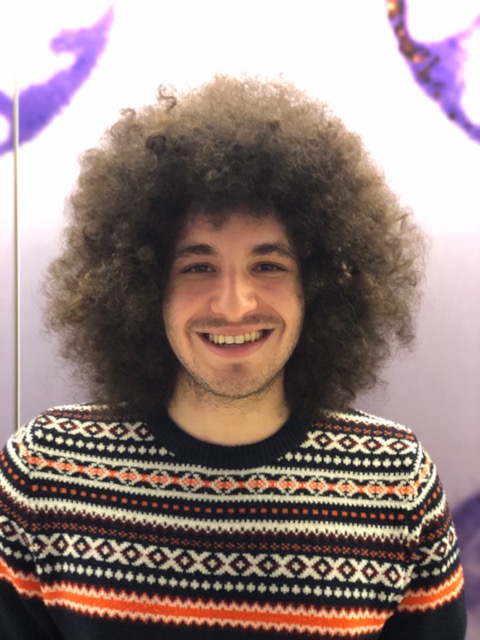
Microscopy and all its uses in biology have fascinated me from the start of my Bachelor’s degree of biology. I was fortunate to receive in-depth training in confocal microscopy during my first internship where I worked on answering research questions in the Zebrafish model. My microscopy training was further expanded during my Master’s degree internship in the Briegel lab. Here I learned how to use cryo-electron microscopy to study bacterial ultrastructure. Combining these two microscopy techniques with the molecular and microbial knowledge that I gained during my Bachelor’s and Master’s degree now allows me to focus on a wide range of research projects. These projects include: investigating the effect of curvature on the polarity of Vibrio cholerae and investigating the use of bacteriophages as a treatment for cholera infected Zebrafish larvae. All of this has been made possible in no small part by the expertise that surrounds me in the Briegel lab and the inspiring environment that I work in!
Janine Liedtke
Graduate Student
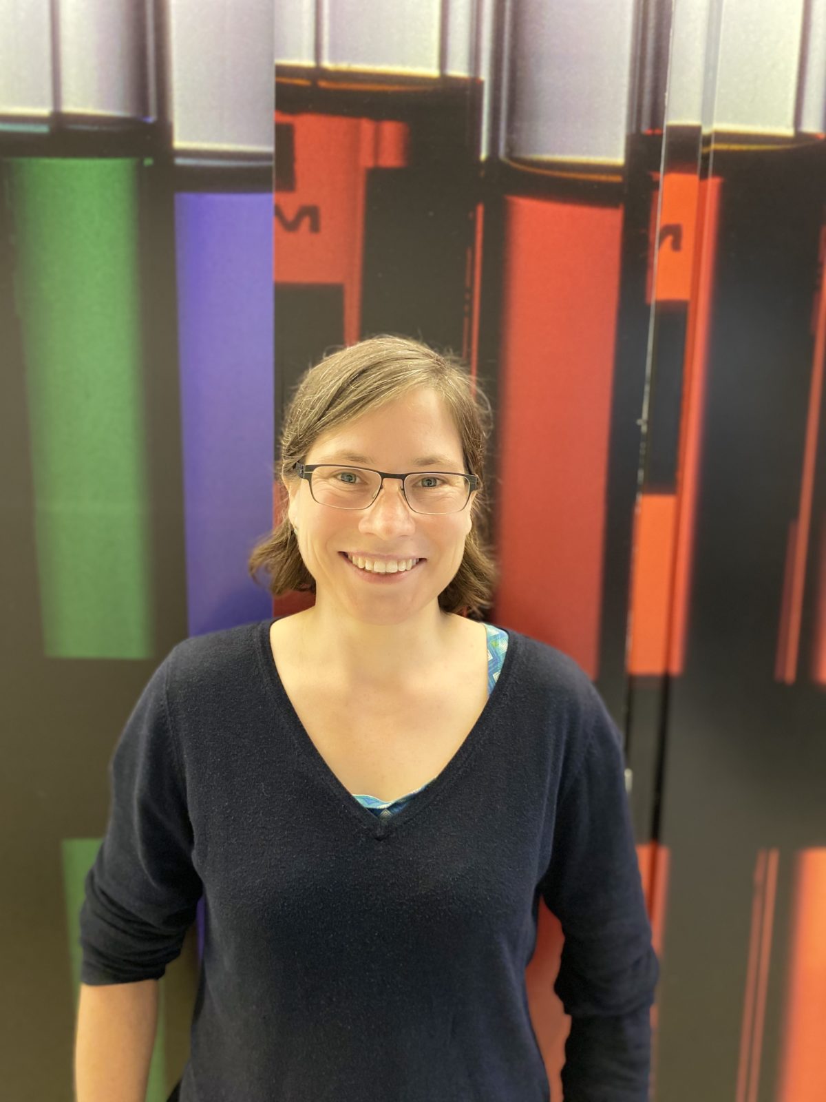
I am fascinated by the fact that a small detail of a system, such a microbe is, can have a tremendous impact and the ability to change a whole system for better or for worse. I want to understand how microbes interact with their environment and what it makes the microbe to change a system in a certain way. After obtaining my degree as medical lab technician and gathering some work experience. I continued my education and obtained a bachelor degree in biology at the Free University of Berlin and a master degree at the University of Bonn. During my whole study I set my focus on microbiology, specifically on bacteria and how they colonize different surfaces as well as how they handle aggravated growth conditions. After obtaining my university degrees I got the great chance to work in interdisciplinary groups at the European space agency, where I among other things supported the work at Human Space flight and Robotics section as well as the artificial photosynthesis project of Dr. Katharina Brinkert. Furthermore, I went to Oslo, Norway, to work on Bacillus cereus spores and their outer surface structure at the Norwegian University of Life Science. During my work on spore structures I learned about cryo-electron microscopy and I was immediately excited about the possibility this technique gives. The Briegel lab gives me the possibility not only to learn the techniques but also to understand how microbes interact and impact their environment. I am currently joining the NWO-groot consortium where I am going to visualize the plant-endophytes interactions by cryo-electron tomography.
Vera Williams
Media Specialist

I am a graduate from the Media Technology master programme from Leiden University. This program taught me to perform my research creatively and to utilise the media platforms that best fit the research. However, my academic roots are grounded in the field of microbiology. These roots were developed during my years in the Biology bachelor program from Leiden University, as well. During the third year of my research internship in the Biology program, I developed an interest in creating 3D models, animations and installations from microbiological data such as microscopy images, which led me unto the unconventional path of scientific visualisations.
Visualising the invisible world of microbes captivated me, and since that moment I have pursued this idea relentlessly. It is my goal to become a proficient scientific visualiser of microbiological topics, and there is still so much I want to discover in this field. But, the quality of these visualisations is heavily dependent on high-quality data and in-depth scientific knowledge on the microbiological topic.
I therefore count myself very lucky to be part of Dr. Ariane Briegel’s lab, as there is plenty to be found of both in her research group. And through Ariane’s close collaboration with NeCEN, the Netherlands Centre of Electron Nanoscopy, the visualisations can be based on the highest resolution microscopy data available today! This allows us to look at microbes on a new, deeper level.
It’s exciting to combine cutting- edge microbiological research with the latest visualisation techniques. Through this, I hope to stimulate communication on microbiological topics among researchers and to the general populace.
Alumni
Katrina Gundlach

Postdoc
My formal science training began in the field. I started my studies at Hawaii Pacific University and received my bachelor’s in Marine Biology in 2012. During these years, I also worked with several different environmental NGOs in Madagascar, Indonesia, and Tanzania as a SCUBA diver. I collected data on the health of marine habitats, helped local governments implement sustainable fishing practices, advised community-led restoration projects, and educated local children and adults on developing sustainable alternative livelihoods. It was from here I decided to go to graduate school and pursue my doctorate. My PhD advisor Dr. Glen Watson at University of Louisiana at Lafayette saw my deep love for Cnidarians and he introduced me to the beautiful world of microscopy. My PhD project looked at the very cool stinging cells of sea anemones and how they are regulated as a function of nutrition and symbiotic state. I finished my PhD in Environmental and Evolutionary Biology in 2018 and started a postdoc in Dr. Margaret McFall-Ngai’s lab at University of Hawaii at Manoa. Here I stated working on the Hawaiian Bobtail Squid. My project looked at how the squid’s ciliated symbiotic organ actively collects its symbiont Vibrio fischeri from the surrounding seawater, using high-speed videography with MATLAB analysis. Now as a postdoc in Dr. Briegel’s lab, I am continuing to use the Squid – Vibrio system to study how the symbiotic bacteria interact with host epithelial cells throughout the development of the symbiotic relationship by using high resolution cryo-EM techniques. I’m very excited to have my squid here in the Netherlands!
Ruochen Ouyang

Graduate Student
I am interested in applying cryoEM to understand the fundamental dynamic processes and mechanisms of antigen-antibody binding and phage infection. I got my bachelor’s degree in physics at Lanzhou University in 2017, then I was directly recommended as a PhD candidate free of examination and studied at Xi’an Jiaotong University. I have learned scientific research skills in the field of CryoEM with professor Lei Zhang. Now I appreciate that I can work in professor Ariane Briegel’s group as a joint training doctoral student.
Alise Muok

Postdoc
My interest in the physical sciences started when I was a student at a community college in California. What first started as a necessary ‘general education’ course in chemistry soon became a personal fascination and career goal. When I transferred to U.C. Davis to earn my bachelor’s degree, I immediately started working in a university biochemistry lab that studies secondary metabolites in plants through biochemical and genetics methods. Fortunately for me, the PI of the lab gave a lot of responsibility and freedom in the lab to undergraduates like myself, which sparked my desire to continue science research at the graduate level. After graduating UC Davis, I started working in the lab or Dr. Brian Crane at Cornell University as a graduate student and was introduced to the world of structural biology. My projects here specifically focused on studying the structure and function of proteins involved in bacterial chemotaxis, primarily through crystallography. Now, in Dr. Ariane’s Briegel’s lab, I am continuing to study bacterial chemotaxis pathways through electron microscopy techniques. As a recipient of the Leading Fellowship for international post-docs, I get the privilege of working in a collaborative environment in a fantastic lab and beautiful country!
Do you want to know more about me? Check out my website!
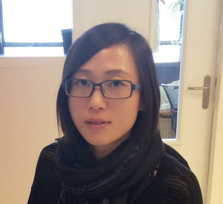
Wen Yang
Graduate Student
I have an interdisciplinary educational background including a bachelor’s degree in pharmacy and a master’s degree in biomedical engineering. Nevertheless, in recent years I have been involved in a series of research projects with a shared focus within life science and biology, from pharmacodynamics studies aiming for target drug release to revealing the molecular mechanism of functional amyloid polymerization in bacteria. In the past two years my primary research focus has been on the “structure and assembly of functional amyloids.” Through this research I have come to realize how exciting, yet challenging, it is to reveal the structure and function of proteins, because this can not be accomplished without a complex set of expertise and techniques and through extensive collaboration across various disciplines.
Eveline Ultee
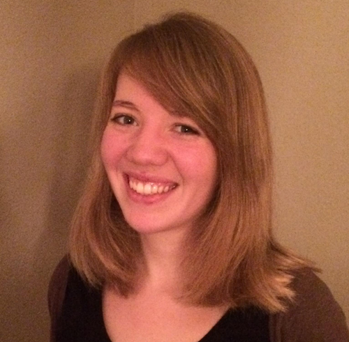
Graduate Student
In the past years I have obtained a Bachelor’s degree and Master’s degree in biology. As a result of my study program, I have gained a broad educational background in research related to health and disease. I got the chance to participate in divers research projects: from a project investigating the effect of oral bacteria on cell proliferation in the human oral cavity, to research focussing on the role of genes in the Campylobacter chemotaxis system. The researchers and experts I have got the opportunity to work with inspired me and gave me an insight into the numerous challenges and possibilities that lay within microbiological research.
Rohola Hosseini

Postdoc
I became interested in the microscopic world and the dynamics in cell biology during my bachelor and master studies at Technical University of Delft and Leiden University. I have finalized my master program in Biophysical Structure Chemistry in the group of Prof. Jan Pieter Abrahams on microtubule dynamics and high-throughput nano-protein crystallization for electron diffraction. Subsequently, I performed my PhD in the group of Prof. Herman Spaink on the role of autophagy during the course of mycobacterial infection in zebrafish larvae model using correlative light and electron microscopy. After my PhD, I have studied Pseudomonas cell physiology in the group of Prof. Han de Winde. Currently, in the group of Prof. Ariane Briegel my research focuses on high-resolution in situ imaging to increase our understanding of Pseudomonas cell physiology and zebrafish host responses during infection.
Susanne Brenzinger
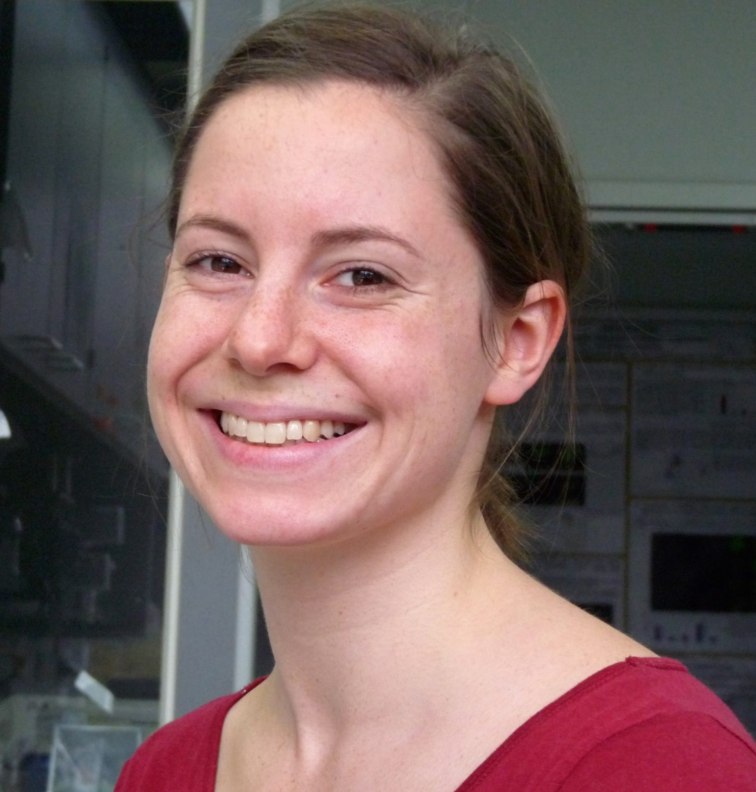
Postdoc
I started my bachelor training in cellular and molecular biology at the University of Osnabrueck, Germany, in 2005 and was immediately drawn to the question of how microbes are capable solving a multitude of tasks with only the tools that are available in one single cell. After a brief excursion into the area of human genetics, I continued my training (now towards the master degree) at the Philipp University of Marburg, Germany, where I joined the lab of Kai Thormann in 2011 to study physiological aspects of the flagellar motor in γ-proteobacteria. For my PhD thesis, I continued to work with Prof. Thormann (since 2013 at the University of Giessen, Germany), now also focusing on questions related to cell polarity and chemotaxis. My PhD studies have further cemented my interest in bacterial cell physiology, which I now seek to understand and analyze in more detail and with higher resolution at the Briegel lab. To do so I received a fellowship in 2017 funded by the German National Academy of Science Leopoldina.
Davi Ortega

Postdoc
I am a computational molecular biophysicist passionate about evolution of complexity and decentralized forms of governments.I am a postdoc at Leiden Universityin the Briegel Lab where I continue my studies in the evolution of chemotaxis networks and data sharing using blockchain technology.I am also one of the developers of the MiST3 database. I received my PhD in Physics with Igor Zhulin at Oak Ridge National Laband University of Tennessee. Next, I spent five years as a postdoctoral scholar at Caltechin the Jensen Labwhere I led the bioinformatics studies in macromolecular machineries and chemotaxis.In a distant past, I worked with Atomic Optical Clocksat the Time and Frequency Division at NISTsupervised by Chris Oatesand Flavio Caldas da Cruz.I am a co-founder of the Art+Science collective Schema47in Los Angeles and I am a member of the core team of the cryptocurrency FLO. I collaborate with the Brazilian Pirate Partyand Vote na Web.
Jamie Depelteau
Postdoc

After receiving my bachelor’s degree in microbiology and cell sciences from the University of Florida, I continued my education at The Ohio State University where I earned a master’s degree in education and completed three years of biomedical science graduate coursework and research. Most recently at the University of California, Berkeley, I had multiple roles including researcher, lecturer, and manager for undergraduate affairs. My research experiences range from genetic and cellular studies of animal models of disease to employing genome editing to generate cell lines that facilitate the study of actin nucleation and how intracellular pathogens hijack this process. My current research interests include understanding the fundamental mechanisms that regulate host-pathogen interactions, specifically how bacteria adjust to changes in the host environment. The Briegel research group and the Institute of Biology provide the tools, expertise, and support to do this with unprecedented clarity.